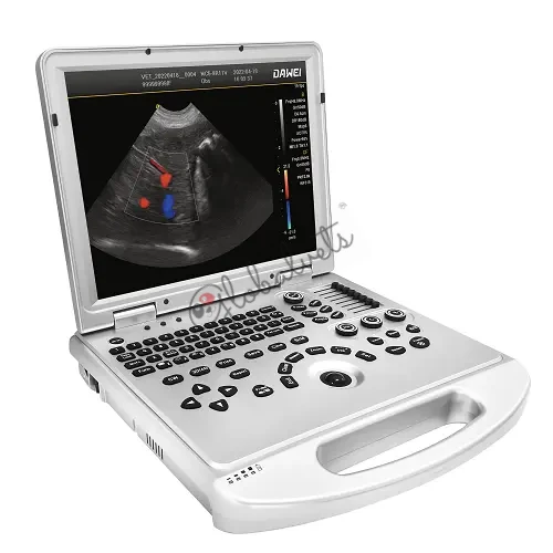
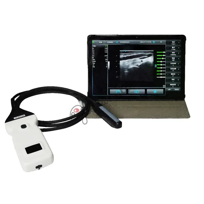
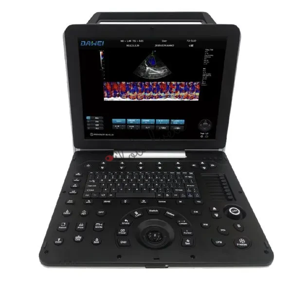
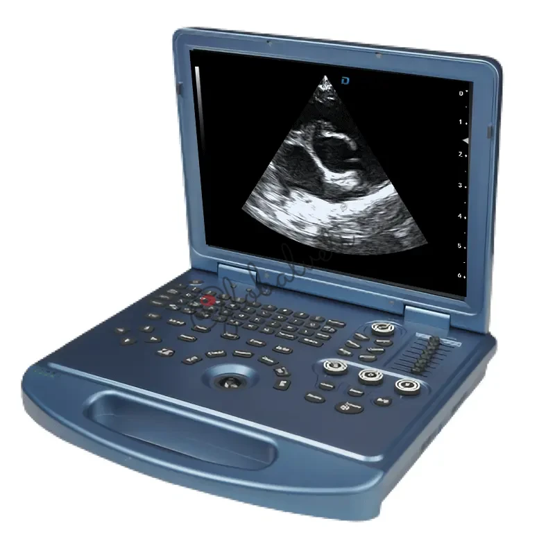
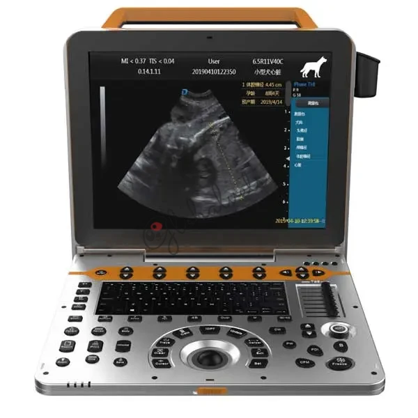
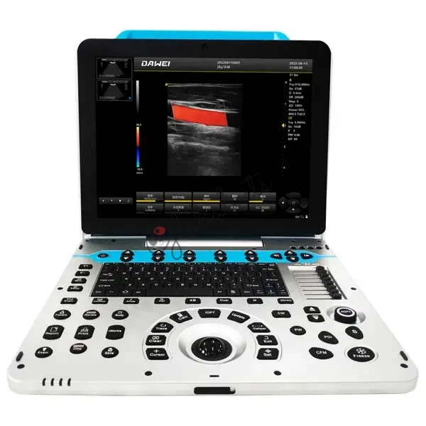
Color Doppler
Enhance your diagnostic capabilities with advanced color Doppler ultrasound, providing high-resolution imaging and precise blood flow analysis for comprehensive cardiovascular and soft tissue examinations.
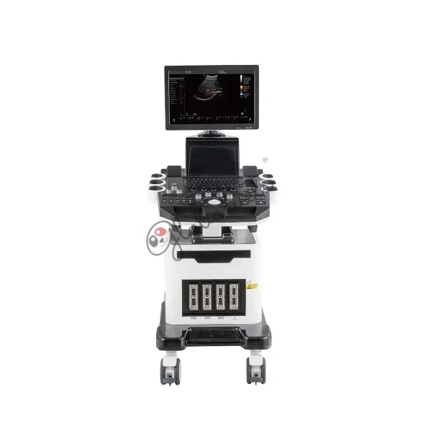
Products
Showing 1-12 of 15 products
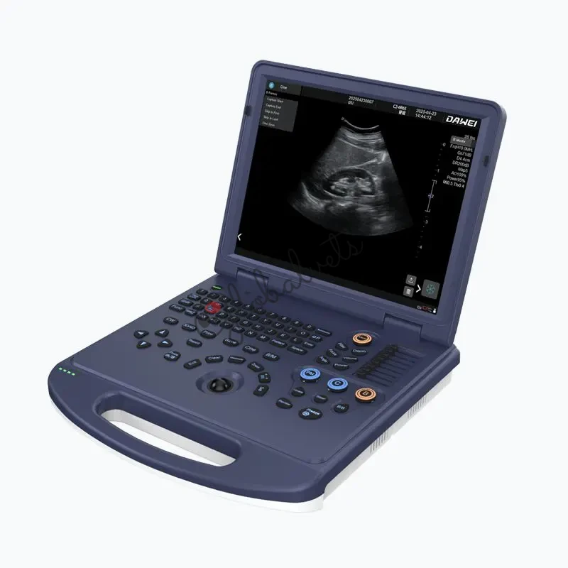

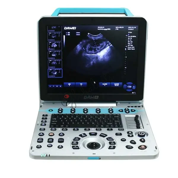
About Color Doppler
Enhance your diagnostic capabilities with advanced color Doppler ultrasound, providing high-resolution imaging and precise blood flow analysis for comprehensive cardiovascular and soft tissue examinations.