Portable Laptop Animal Reptile Ultrasound Machine Veterinary Ultrasound Scanner
Portable laptop veterinary ultrasound scanner can be used for the examination of different organs in sheep, dogs, cats, reptiles, etc. The images are preset to optimize the examination conditions and reduce the adjustment during veterinary operation.
Need Expert Advice?
Call us: +91 9811520889
Applications
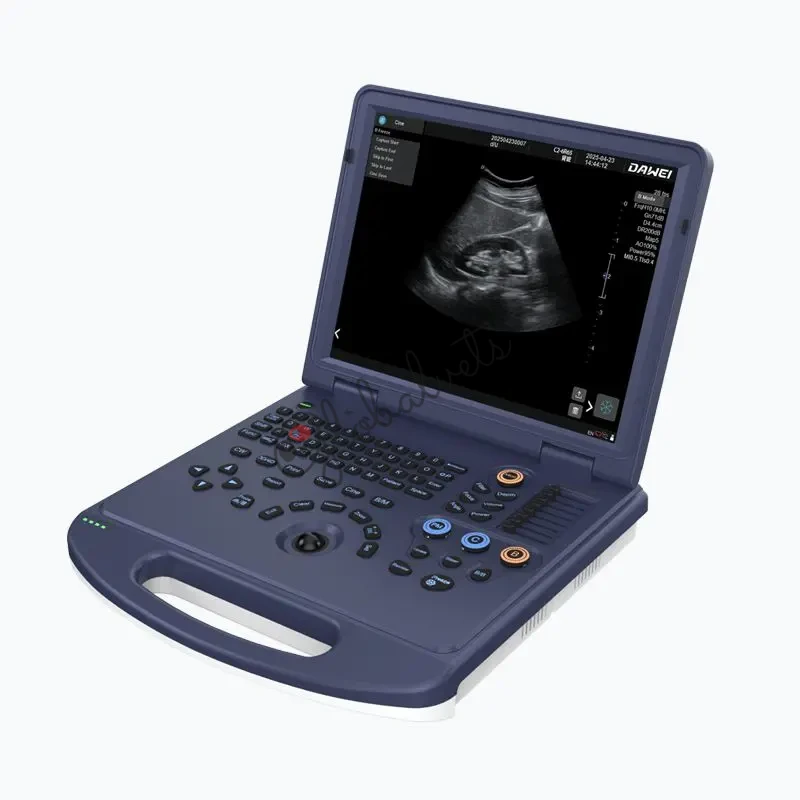
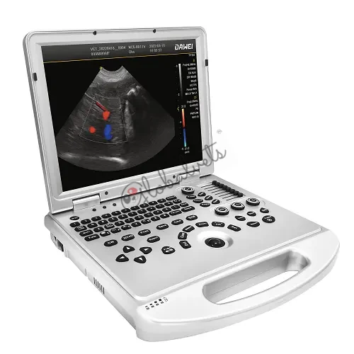
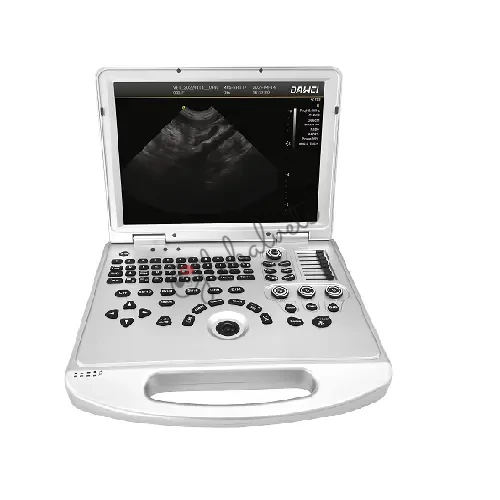
The L3-VET Veterinary Color Doppler Ultrasound System is a portable laptop-style veterinary ultrasound scanner designed for daily imaging needs in pet hospitals, veterinary clinics, zoos, breeding/reproduction bases, and research institutions.
It combines B-mode, Color Doppler, and Spectral (PW) Doppler with practical image optimization features for fast workflow and confident diagnosis.
Target animals: companion animals and exotics (including reptiles, depending on probe selection and clinical protocol).
For the diagnosis of reptile pregnancy, diagnosis of fish gender.
Key Benefits (Why Clinics Choose L3-VET)
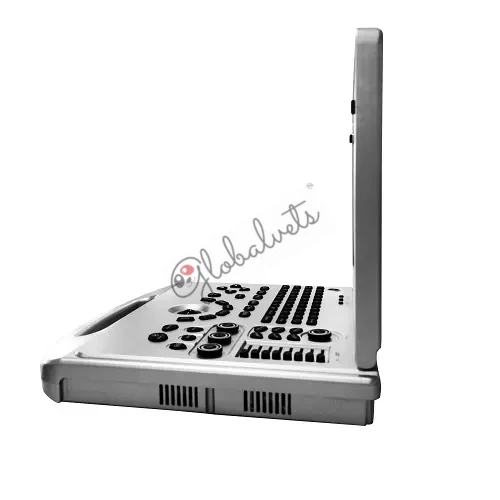
- Laptop-type portability for multi-room clinics, mobile services, and limited spaces
- Color Doppler + PW Doppler to evaluate blood flow and hemodynamics
- Image enhancement tools: THI (Tissue Harmonic Imaging), Compound Imaging, Trapezoidal Imaging
- One-Key Smart Optimization to reduce manual adjustments
- 15-inch monitor for clear viewing during fast-paced exams
- All-in-one clipboard workflow: thumbnails displayed at the bottom for quick save / transfer / delete
- DICOM 3.0 support for PACS connectivity and standardized image management
Imaging Modes & Technologies
Core Modes
- B / C / D real-time three synchronous imaging
- PW (Pulsed Wave) Doppler
- DPDI (Directional Power Doppler Imaging)
- 2B / 4B imaging modes
- B/C split screen (real-time)
Image Quality & Efficiency
- THI (Tissue Harmonic Imaging)
- Compound Imaging
- Trapezoidal Imaging
- One-Key Smart Optimization
- Preset exam conditions for different organs (reduces repeated adjustments)
2D (B-Mode)
| Parameter | Range / Levels |
| Gain | 0–100 (step 1) |
| TGC | 8 segments |
| Dynamic range | 20–280 (20 levels) |
| Pseudo color | 0–11 |
| Sound power | 5%–100% (step 5%) |
| Max focus | ≥ 6 |
| Line density | low / middle / high |
| Noise reduction | 0–14 |
Color Doppler
| Parameter | Range / Levels |
| Color frame correlation | 0–12 |
| Color map | 0–7 |
| Baseline | 11 levels |
| Line density | low / high |
| Filter | 0–5 |
Spectral (PW) Doppler
| Parameter | Range / Levels |
| Angle correction | -80° to 80° |
| Sample volume | 0.5–20 mm |
| Speed scale | 32.8–328 cm/s (varies by probe) |
| Auto calculations | PS, ED, RI, PI, S/D, HR, etc. |
Clinical Applications
Suitable for ultrasound examination needs in:
- Pet hospitals and veterinary clinics
- Zoos and wildlife/exotic animal facilities
- Breeding and reproduction bases
- Universities and scientific research units
Common veterinary use cases include:
- Abdomen (liver, kidney, spleen, bladder, etc.)
- Reproductive evaluation (OB)
- General soft-tissue scanning
- Doppler vascular assessment (as clinically required)
Parameter Control Highlights
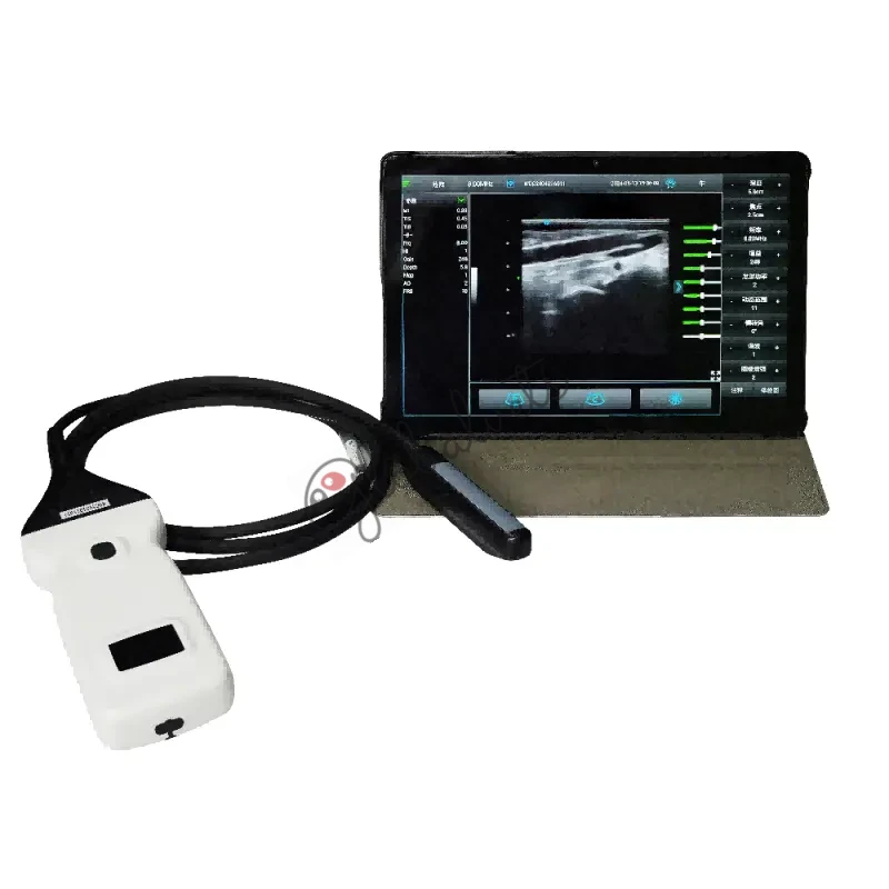
Probe
Probe interface ≥ 1
Final probe configuration depends on your quotation and local regulatory requirements.
| Probe Type | Detecting Depth | Fundamental Frequency | Harmonic Frequency |
| Convex | 30–255 mm | 2.0 / 2.5 / 3.0 / 3.5 / 4.0 / 5.5 MHz | H4.0 / H5.0 MHz |
| Linear | 20–128 mm | 6.0 / 7.5 / 8.5 / 10.0 / 12.0 MHz | H10.0 MHz |
| Micro-convex (R11) | 30–111 mm | 4.5 / 5.0 / 6.0 / 6.5 / 7.0 / 9.0 MHz | H8.0 MHz |
| Rectal | 20–111 mm | 4.0 / 6.5 / 9.0 MHz | H8.0 MHz |
![]()
Quick Specifications
| Item | Specification |
| System type | Veterinary Color Doppler ultrasound diagnostic system |
| Form factor | Laptop type |
| Operating system | Windows 10 |
| Display | 15-inch main monitor |
| Doppler | Color Doppler, DPDI (Directional Power Doppler Imaging), PW (Pulsed Wave Doppler) |
| Imaging sync | B / C / D real-time three synchronous imaging |
| Advanced imaging | THI, Compound Imaging, Trapezoidal Imaging |
| Multi-frame modes | 2B / 4B |
| Presets | Preset exam conditions for different organs |
| Upgrade | On-the-spot upgrade function |
| Probe ports | 1 probe interface |
| Storage | Built-in 128GB hard disk |
| Image formats | BMP / DCM / JPG |
| Movie playback | ≥ 600 frames |
| DICOM | DICOM 3.0, can connect to PACS |
| Interfaces | 4×USB, 1×Audio, 2×RJ-45, 1×HDMI |
| System languages | Chinese / English / French / Russian / Spanish |
Connectivity & Interfaces
| Interface | Quantity |
| USB | 4 |
| Audio | 1 |
| RJ-45 (LAN) | 2 |
| HDMI | 1 |
Measurement, Reporting & Data Management
- General measurements: distance, area, angle, time, slope, HR, speed, acceleration, flow parameters, etc.
- Professional data packages for different organs (examples): Abdomen, OB
- Built-in patient/file management: ID, name, exam number, exam date, search and management
- One-key fast report function
Standard Configuration & Optional Accessories
Standard configuration
- Full digital veterinary color Doppler ultrasound diagnostic system (main unit)
Optional
- Probes: R11 micro-convex / convex / linear / rectal
- Video printer
- Trolley
You May Also Like
Explore similar products for your veterinary practice