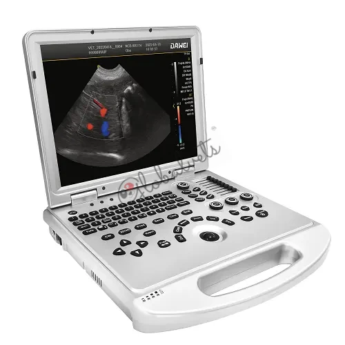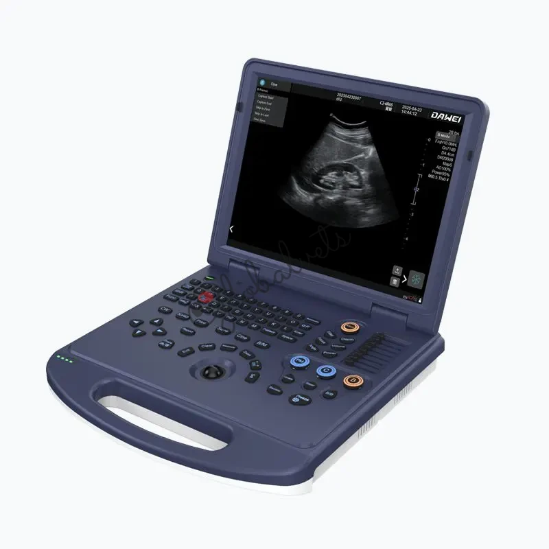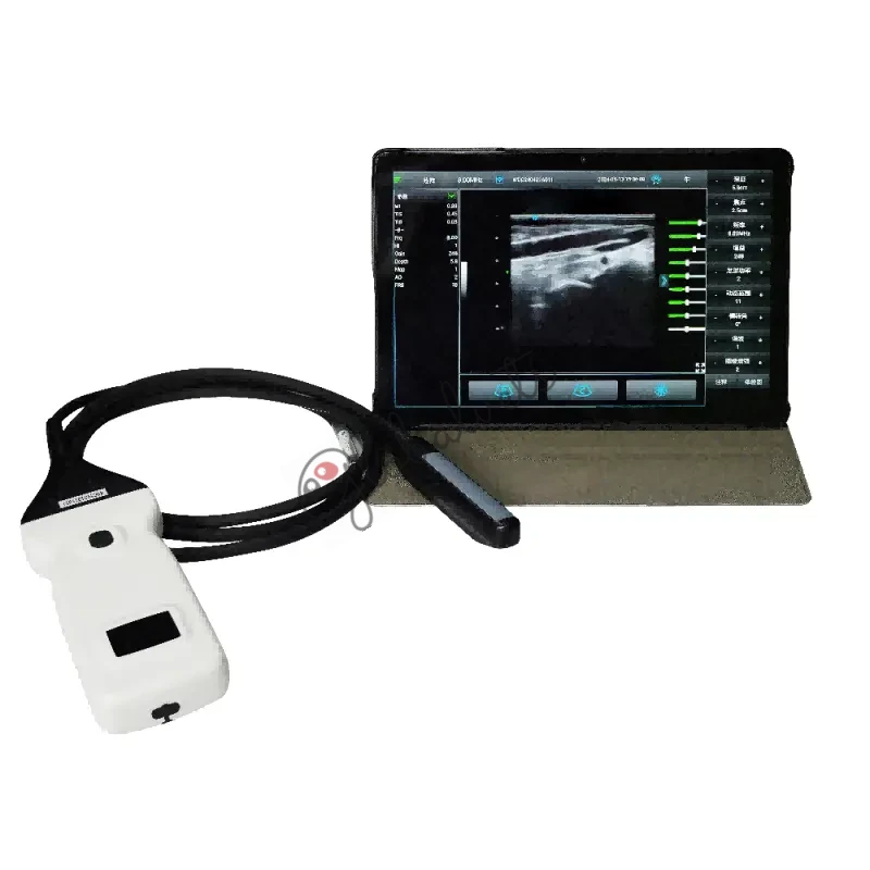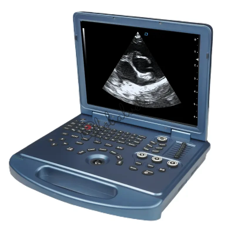
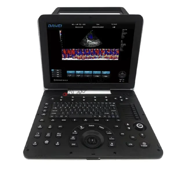
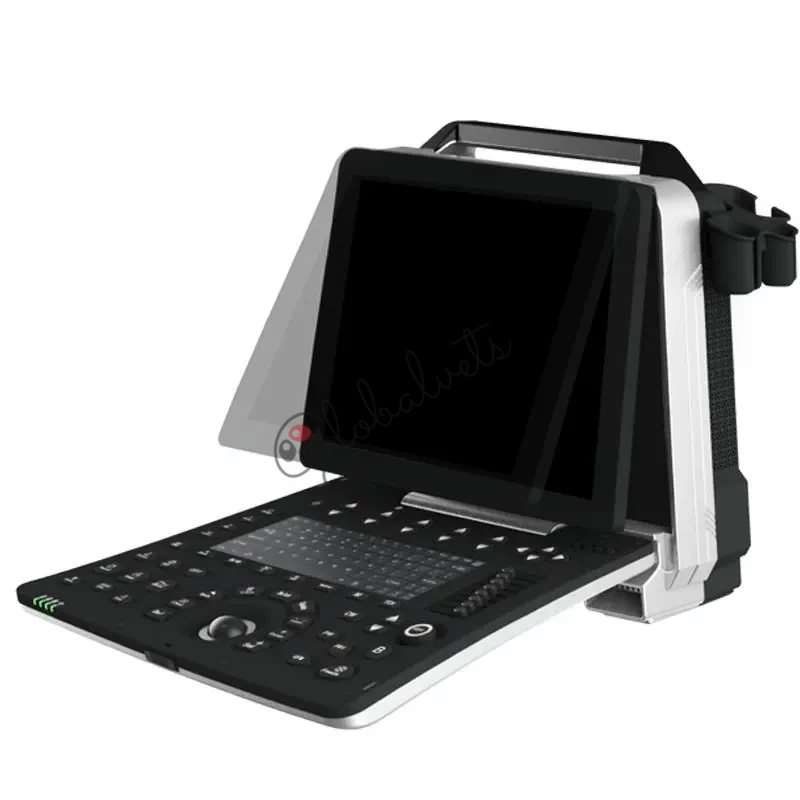
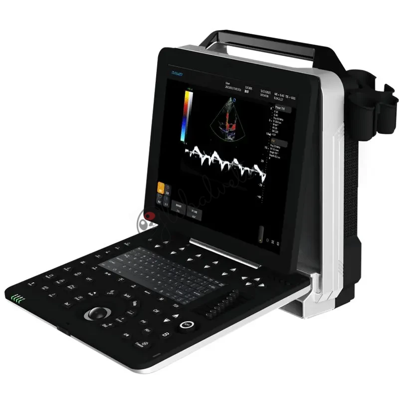
Portable Veterinary Doppler Ultrasound Machine Imaging for Dogs, Cats & Exotic Animals
The P5S-VET Animal Color Doppler Ultrasound System is a premium portable veterinary ultrasound machine built for pet clinics, animal research centers, and mobile veterinary diagnostics. Designed with superior Doppler imaging technology, ergonomic portability, and advanced GPU-enhanced computing, it provides fast, precise diagnostic results in a compact, cost-effective solution.
Need Expert Advice?
Call us: +91 9811520889
Applications
Companion animal diagnostics (dogs, cats, rabbits, exotics)
Veterinary hospitals and specialty clinics
Mobile veterinary services, ICU, anesthesia, and emergency diagnostics
Academic & animal research institutions
Whether you're conducting cardiovascular imaging, abdominal scanning, or musculoskeletal evaluations, the P5S-VET pet ultrasound scanner is engineered to perform with precision and reliability in various clinical settings.
Performance
High-speed parallel computing using CPU + GPU dual-core architecture
Up to 2x faster frame rate, 10x faster response, and enhanced real-time rendering
Seamless real-time B/C/D mode, PW/CW Doppler, TDI, and M-color flow imaging
ST-U Imaging Platform with CPU+GPU Acceleration
Optimized System Features
15" full-HD medical-grade screen with 0–30° tilt adjustment
Windows 7 platform with user-friendly interface
256GB SSD storage, DICOM support, HDMI output
USB 3.0 for fast export of images, videos, and reports
Internal battery: ≥2 hours of operation for mobile vet use
Advanced Probe & Imaging Capabilities
Imaging Modes & Diagnostic Tools
Tissue Harmonic Imaging (THI) & Frequency Compound Imaging
Elasticity Imaging to differentiate soft vs. hard tissue
OmniView free-plane imaging for complex structures
Wide Field of View (PView) for large lesions and panoramic anatomy
Speckle Noise Reduction, Auto Doppler Envelope Tracking, and more
Probe
Linear and convex probes available for different animal sizes and organ depths
Advanced beamforming + A/D conversion = high-resolution images
Needle enhancement for precise interventional procedures
You May Also Like
Explore similar products for your veterinary practice